Ankle Arthroscopy
People use eye-hand coordination all the time to perform daily tasks and activities—like typing on a computer keyboard while watching the monitor, playing piano while reading sheet music, or swinging a baseball bat while keeping an eye on the ball. Modern medicine has applied the same principle to how we do surgery. Now, instead of bending over a table to see into an incision and watch hand movements, surgeons often watch a picture on a television screen while manipulating a camera and tools from outside the foot. Welcome to the world of ankle arthroscopy!
How Ankle Arthroscopy Works
 When you need surgery for an ankle problem—say to remove a bone spur or repair cartilage or ligament damage—we use a metal tube a little smaller than a pencil called an arthroscope that is inserted through a tiny incision in the skin into the joint capsule. This is a watertight pocket around the ankle joint made up of soft tissues like tendons, ligaments, and membranes that is filled with sterile saline solution during the procedure. You might think of it like a drinking straw inserted into a water balloon.
The fiber-optics that run through the tube have a camera, light, and tiny tools on the ends of them. The camera sends pictures back through the cables to a screen standing next to the surgeon. This is what he looks at as he turns on the light, examines the area inside the joint, and performs the surgery.
For ankle arthroscopy, you will be on your back with your leg bent and lifted and the affected ankle positioned and held in place with traction. This slightly separates the bones to allow the tube to be inserted. You will be under general anesthesia (asleep) during the procedure. Small incisions are made for the arthroscope, a cannula to inject the saline, and the miniscule tools needed to do the procedure(s).
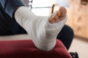
When the surgery is over, everything is removed, the capsule and skin incisions stitched or stapled, and dressings and compression stockings or splints applied.
What Minimally Invasive Surgery Is Used For
We mentioned a couple of conditions benefiting from this type of surgery—removing bone spurs or repairing a ligament or cartilage. This method can also be used to confirm a diagnosis, remove loose fragments in the joint complex, or get rid of scar tissue or inflamed tissue that is causing problems. It can even be used to repair ankle fractures and treat ankle arthritis in some cases.
Because the images are magnified on the screen, it is often easier for us to see where the damage is and what needs to be done. Since the incision is small, there is less tissue damage from the procedure, and recovery times are usually shortened.
As with any surgery, there will be risks of anesthesia complications, clotting, infection, and failure of equipment, but we will discuss everything with you beforehand so you know what to expect from the surgery and recovery.
Help from Southern California Foot & Ankle Surgeons
Dr. Robert Spencer and Dr. Nitza Rodriguez are experienced ankle surgeons who can help you with ankle pain. Come in and let us evaluate what’s going on and offer treatment options for your consideration. Our goal is to get your ankles into as healthy a state as possible so you can enjoy your life without pain or limitation.
Call Southern California Foot & Ankle Specialists today at (949) 364-9255 (WALK) or schedule an appointment online and learn what we can do for you.
Contact Us
Ladera Ranch
333 Corporate Dr. Ste 230, Ladera Ranch, CA 92694
Tel: (949) 364-9255 (WALK)
Fax: (949) 364-9250
Office Hours:
Monday - Friday: 9am - 5pm
*(Lunch 12 noon - 1pm)
Orange
2617 E Chapman Ave. Ste 303, Orange, CA 92869
Tel: (714) 639-7993
Fax: (714) 639-0729
Office Hours:
Monday - Friday: 9am - 5pm
*(Lunch 12 noon - 1pm)